书馨卡帮你省薪
欢迎光临中图网 请 | 注册
> >>
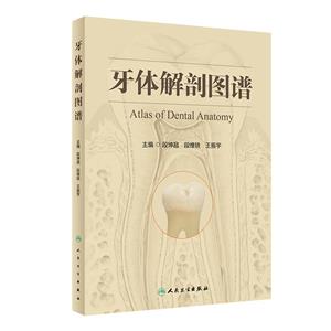
牙体解剖图谱

牙体解剖图谱

出版社:人民卫生出版社出版时间:2024-02-01
开本: 16开 页数: 240
本类榜单:医学销量榜
中 图 价:¥141.0(7.5折) 定价  ¥188.0 登录后可看到会员价
加入购物车 收藏
运费6元,满39元免运费
?新疆、西藏除外
本类五星书更多>

牙体解剖图谱 版权信息

  • ISBN:9787117341998
  • 条形码:9787117341998 ; 978-7-117-34199-8
  • 装帧:一般铜版纸
  • 册数:暂无
  • 重量:暂无
  • 所属分类:>>

牙体解剖图谱 本书特色

始终坚持以真实的形态、典型的结构为基础,以适用于口腔医学教学为重点,对口腔颌面部的各器官从巨视解剖和微视解剖的正常形态结构与临床有关的变异、畸形、病理改变进行了较为系统而全面的展示。 内容翔实、准确、联系实际;图像清晰、真实、鲜明易懂。

牙体解剖图谱 内容简介

采用成年人恒牙、牙列、咬合、牙合曲线、牙合型及婴幼儿乳牙、牙列、咬合、牙合曲线、牙合型的实物标本。观察牙体的各面结构及牙体的各种剖面的特征,力求将每个牙体微小结构、形态真实的展现读者面前。该图谱强化结构的标注和文字描述,如每个牙重要结构名称,临床应用意义,重要要点进行文字说明,做到图文并茂,达到牙体解剖学的认知充分服务于口腔临床目的,弥补各院校口腔教学中牙体标本奇缺,多采用教学模型的弊端。使学生能够轻松的掌握教材中的牙体解剖学知识和内容。图像按口腔解教材内容的顺序编排,用栩栩如生的牙体实物标本图像,来阐述死记硬背的牙体解剖学知识,并对每个牙的解剖特征、牙齿的要素、牙体的变异、牙髓腔、根管的形态等进行词条性文字描述。图解随图同伴,方便便捷。

牙体解剖图谱 目录

**章 口腔颌面部的标志/1 Marker of oral cavity and maxillofacial region 图1 颅的概观(前面观)General view of skull. Anterior view/2 图2 颅的概观(侧面观)General view of skull. Lateral view/3 图3 老年颅的概观(前面观)General view of old skull. Anterior view/4 图4 老年颅的概观(侧面观)General view of old skull. Lateral view/5 图5 乳牙的分类与命名Name and grouping of deciduous teeth/6 图6 恒牙分类与命名Name and grouping of permanent teeth/6 图7 乳牙的名称和符号Name and denotation of deciduous teeth/7 图8 恒牙的名称和符号Name and denotation of permanent teeth/8 图9 牙的组成部分Ingredient of tooth/8 图10 牙的构造示意图Diagram of structure of tooth/9 图11 牙髓腔的示意图Diagram of dental pulp cavity/11 图12 根管的分型示意图Diagram of grouping of root canal/12 图13 根管与根尖孔变异示意图Diagram of variation of root canal and apical foramen/14 图14 牙本质和牙釉质的形态Shapes of dentin and enamel/15 图15 牙体长轴Long axis of tooth/16 图16 牙体外形高点Height of contour of tooth/17 图17 牙邻面接触区部位Site of contact area of proximal surface of tooth/18 图18 牙邻面及面接触区部位Site of occlusal contact area and proximal surface of tooth/18 图19 牙冠的线角Line angle of dental crown/19 图20 牙冠的点角Point angle of dental crown/20 图21 切牙牙体三等分Division into thirds of incisor teeth/21 图22 磨牙牙体三等分Division into thirds of molar/21 图23 牙冠各面的名称Name of each surfaces of dental crown/22 图24 上颌牙冠的各面Each surfaces of dental crown of maxilla/23 图25 下颌牙冠的各面Each surfaces of dental crown of mandible/24 图26 牙冠的表面标志Surface marker of dental crown/25 图27 牙尖Dental cusp/26 图28 切缘结节Mamelon/26 图29 舌面隆凸Cingulum/27 图30 切嵴Incisal ridge/27 图31 轴嵴和颈嵴Axial ridge and cervical ridge/28 图32 边缘嵴Marginal ridge/29 图33 牙尖嵴Cusp ridge/29 图34 三角嵴Triangular ridge/30 图35 横嵴Transverse ridge/30 图36 斜嵴Oblique ridge/31 图37 发育沟、点隙、副沟Developmental groove、pit、supplemental groove/31 图38 裂Fissure/32 图39 窝Fossa/32 图40 斜面Inclined surface/33 图41 牙冠的生长叶Growth lobe of crowns/34 图42 牙冠接触区的楔状隙Embrasure of contact area of crowns/35 图43 牙邻面接触区的位置Site of proximate contact area of tooth/36 第二章 恒牙与牙髓腔的形态/39 图44 上颌切牙示意图Diagram of incisor of maxilla/40 图45 上颌中切牙Central incisor of maxilla/42 图46 上颌中切牙牙冠的形态Shapes of dental crown of central incisor of maxilla/42 图47 上颌中切牙变异Variation of central incisor of maxilla/43 图48 上颌中切牙牙髓腔的形态Shapes of dental pulp cavity of central incisor of maxilla/43 图49 上颌中切牙髓室顶的形态Shapes of roof of pulp chamber of central incisor of maxilla/44 图50 上颌侧切牙Lateral incisor of maxilla/45 图51 上颌侧切牙(双根)Lateral incisor of maxilla.Double roots/46 图52 上颌侧切牙变异Variation of lateral incisor of maxilla/47 图53 上颌侧切牙牙髓腔的形态Shapes of dental pulp cavity of lateral incisor of maxilla/47 图54 上颌尖牙示意图Diagram of canine tooth of maxilla/48 图55 上颌尖牙Canine tooth of maxilla/49 图56 上颌尖牙(短根)Canine tooth of maxilla.Short root/50 图57 上颌尖牙变异Variation of canine tooth of maxilla/51 图58 上颌尖牙阻生及错位Impaction and transposition of canine tooth of maxilla/51 图59 上颌尖牙牙髓腔的形态Shapes of dental pulp cavity of canine tooth of maxilla/52 图60 上颌**前磨牙示意图Diagram of 1st premolar of maxilla/53 图61 上颌**前磨牙(双根)1st premolar of maxilla.Double roots/54 图62 上颌**前磨牙(单根)1st premolar of maxilla.Simple root/55 图63 上颌**前磨牙牙髓腔的形态Shapes of dental pulp cavity of 1st premolar of maxilla/56 图64 上颌第二前磨牙示意图Diagram of 2nd premolar of maxilla/57 图65 上颌第二前磨牙(单根)2nd premolar of maxilla.Single root/59 图66 上颌第二前磨牙(双根)2nd premolar of maxilla.Double roots/60 图67 上颌第二前磨牙牙髓腔的形态Shapes of dental pulp cavity of 2nd premolar of maxilla/61 图68 上颌**磨牙示意图Diagram of 1st molar of maxilla/62 图69 上颌**磨牙(三根)1st molar of maxilla.Three roots/64 图70 上颌**磨牙(三根融合)1st molar of maxilla.Three roots fusion/65 图71 上颌**磨牙牙髓腔的形态Shapes of dental pulp cavity of 1st molar of maxilla/67 图72 上颌第二磨牙示意图Diagram of 2nd molar of maxilla/68 图73 上颌第二磨牙(三根)2nd molar of maxilla.Three roots/69 图74 上颌第二磨牙(双根1)2nd molar of maxilla.Double root(1)/70 图75 上颌第二磨牙(双根2)2nd molar of maxilla.Double roots(2)/71 图76 上颌第二磨牙(尖形双根)2nd molar of maxilla.Apiciform double roots/72 图77 上颌第二磨牙(尖形单根)2nd molar of maxilla.Simple root of apiciform/73 图78 上颌第二磨牙(三根融合)2nd molar of maxilla.Fusion of three roots/74 图79 上颌第二磨牙(近远中颊根与舌根融合)2nd molar of maxilla.Fusion of mesial distal buccal root and lingual root/75 图80 上颌第二磨牙面的形态Shapes of occlusal surface of 2nd molar of maxilla/76 图81 上颌第二磨牙牙髓腔的形态Shapes of dental pulp cavity of 2nd molar of maxilla/77 图82 上颌第三磨牙(三根)3rd molar of maxilla.Three roots/78 图83 上颌第三磨牙(双根)3rd molar of maxilla.Double roots/79 图84 上颌第三磨牙(单根)3rd molar of maxilla.Single root/80 图85 上颌第三磨牙(四根)3rd molar of maxilla.Four roots/81 图86 上颌第三磨牙面的形态Shapes of occlusal surface of 3rd molar of maxilla/81 图87 右上颌第三磨牙变异Variation of 3rd molar of right maxilla/82 图88 上颌第三磨牙牙髓腔的形态Shapes of dental pulp cavity of 3rd molar of maxilla/83 图89 下颌中切牙Central incisor of mandible/84 图90 中切牙与侧切牙(融合牙)Fusion of central incisor and lateral incisor/85 图91 下颌中切牙先天性缺如Congenital absence of central incisor of mandible/86 图92 下颌中切牙牙髓腔的形态Shapes of dental pulp cavity of central incisor of mandible/86 图93 下颌侧切牙Lateral incisor of mandible/87 图94 下颌侧切牙牙髓腔的形态Shapes of dental pulp cavity of lateral incisor of mandible/88 图95 下颌尖牙Canine tooth of mandible/89 图96 下颌尖牙牙髓腔的形态Shapes of dental pulp cavity of canine tooth of mandible/90 图97 下颌**前磨牙示意图Diagram of 1st premolar of mandible/91 图98 下颌**前磨牙1st premolar of mandible/93 图99 下颌**前磨牙(根分叉痕迹)1st premolar of mandible.Vestige of root bifurcation/94 图100 下颌**前磨牙(双根)1st premolar of mandible.Double roots/95 图101 下颌**前磨牙牙髓腔的形态Shapes of dental pulp cavity of 1st premolar of mandible/96 图102 下颌第二前磨牙示意图Diagram of 2nd premolar of mandible/97 图103 下颌第二前磨牙2nd premolar of mandible/98 图104 下颌第二前磨牙面的形态Shapes of occlusal surface of 2nd premolar of mandible/98 图105 下颌第二前磨牙牙髓腔的形态Shapes of dental pulp cavity of 2nd premolar of mandible/99 图106 下颌**磨牙示意图Diagram of 1st molar of mandible/100 图107 下颌**磨牙(三根)1st molar of mandible.Three roots/101 图108 下颌**磨牙(双根)1st molar of mandible.Double roots/102 图109 下颌**磨牙(远中舌根)1st molar of mandible.Distal lingual root/103 图110 下颌**磨牙牙髓腔的形态Shapes of dental pulp cavity of 1st molar of mandible/104 图111 下颌第二磨牙示意图Diagram of 2nd molar of mandible/105 图112 下颌第二磨牙(双根)2nd molar of mandible.Double roots/106 图113 下颌第二磨牙(三根)2nd molar of mandible.Three roots/107 图114 下颌第二磨牙(“C”形根)2nd molar of mandible.“C”root/108 图115 下颌第二磨牙牙髓腔的形态Shapes of dental pulp cavity of 2nd molar of mandible/109 图116 下颌第三磨牙(三根)3rd molar of mandible.Three roots/110 图117 下颌第三磨牙(双根)3rd molar of mandible.Double roots/111 图118 下颌第三磨牙(尖根)3rd molar of mandible.Apical root/112 图119 下颌第三磨牙(融合根)3rd molar of mandible.Fusion root/112 图120 下颌第三磨牙(四根)3rd molar of mandible.Four roots/113 图121 下颌第三磨牙(多根尖)3rd molar of mandible.Multi-root tip/114 图122 下颌第三磨牙釉珠Enamel pearl of 3rd molar of mandible/114 图123 智齿横生Amphitropic wisdom teeth/115 图124 下颌第三磨牙面形态Shapes of occlusal surface of 3rd molar of mandible/116 图125 下颌第三磨牙牙髓腔的形态 Shapes of dental pulp cavity of 3rd molar of mandible/117 第三章 乳牙与牙髓腔的形态/119 图126 上颌乳中切牙Deciduous central incisor of maxilla/120 图127 上颌乳中切牙牙髓腔的形态Shapes of dental pulp cavity of deciduous central incisor of maxilla/121 图128 上颌乳侧切牙Deciduous lateral incisor of maxilla/122 图129 上颌乳侧切牙牙髓腔的形态Shapes of dental pulp cavity of deciduous lateral incisor of maxilla/123 图130 上颌乳尖牙Deciduous canine tooth of maxilla/124 图131 上颌乳尖牙牙髓腔的形态Shapes of dental pulp cavity of deciduous canine tooth of maxilla/125 图132 上颌**乳磨牙1st deciduous molar of maxilla/126 图133 上颌**乳磨牙牙髓腔的形态Shapes of dental pulp cavity of 1st deciduous molar of maxilla/127 图134 上颌第二乳磨牙2nd deciduous molar of maxilla/128 图135 上颌第二乳磨牙牙髓腔的形态Shapes of dental pulp cavity of 2nd deciduous molar of maxilla/129 图136 下颌乳中切牙Deciduous central incisor of mandible/130 图137 下颌乳中切牙牙髓腔的形态Shapes of dental pulp cavity of deciduous central incisor of mandible/131 图138 下颌乳侧切牙Deciduous lateral incisor of mandible/132 图139 下颌乳侧切牙牙髓腔的形态Shapes of dental pulp cavity of deciduous lateral incisor of mandible/133 图140 下颌乳尖牙Deciduous canine tooth of mandible/134 图141 下颌乳尖牙牙髓腔的形态Shapes of dental pulp cavity of deciduous canine tooth of mandible/135 图142 下颌**乳磨牙1st deciduous molar of mandible/136 图143 下颌**乳磨牙牙髓腔的形态Shapes of dental pulp cavity of 1st deciduous molar of mandible/137 图144 下颌第二乳磨牙2nd deciduous molar of mandible/138 图145 下颌第二乳磨牙牙髓腔的形态Shapes of dental pulp cavity of 2nd deciduous molar of mandible/139 第四章 牙列与/141 Dentition and occlusion 图146 恒牙上牙列( 面观)Superior dentition of permanent occlusion. Occlusal surface view/142 图147 恒牙下牙列( 面观)Inferior dentition of permanent occlusion. Occlusal surface view/142 图148 乳牙上牙列( 面观)Superior dentition of deciduous occlusion.Occlusal surface view/143 图149 乳牙下牙列( 面观)Inferior dentition of the deciduous occlusion. Occlusal surface view/143 图150 乳牙上牙列(前面观)Superior dentition of deciduous occlusion. Anterior view/144 图151 乳牙下牙列(前面观)Inferior dentition of deciduous occlusion.Anterior view/144 图152 恒牙上牙列(侧面观)Superior dentition of permanent occlusion.Lateral view/145 图153 乳牙上牙列(侧面观)Superior dentition of deciduous occlusion.Lateral view/145 图154 混合牙列Mixed dentition/146 图155 牙列的形态Shapes of dentition/147 图156 牙列长度和宽度Length and breadth of dentition/148 图157 异常牙列(牙数异常)Abnormity dentition.Numbers abnormity/149 图158 异常牙列(牙排列异常)Abnormity dentition.Malalinement/149 图159 恒牙(前面观)Permanent occlusion.Anterior view/150 图160 恒牙(侧面观)Permanent occlusion.Lateral view/150 图161 乳牙(前面观)Deciduous occlusion.Anterior view/151 图162 乳牙(侧面观)Deciduous occlusion.Lateral view/151 图163 平面Occlusal plane/152 图164 下颌牙列纵曲线Sagittal curve of occlusion of dentition of mandible/152 图165 上颌牙列的纵曲线Sagittal curve of occlusion of dentition of maxilla/153 图166 上颌横曲线Transverse curve of occlusion of maxilla/153 图167 下颌横曲线Transverse curve of occlusion of mandible/154 图168 Bonwill 三角Bonwill triangle/154 图169 Balkwil 角Balkwil angle/155 第五章 咬合/157 Occlusion 图170 牙尖交错牙的接触关系(前面观)Contact relation of malocclusion tooth. Anterior view/158 图171 牙尖交错牙的接触关系(侧面观)Contact relation of malocclusion tooth. Lateral view/158 图172 牙尖交错牙的接触关系(内侧面观)Contact relation of malocclusion tooth.Medial view/159 图173 上颌牙的倾斜方向(前面观)Tilted direction of maxillary teeth. Anterior view/160 图174 上颌牙的倾斜方向(外侧面观1)Tilted direction of maxillary teeth. Lateral view(1)/160 图175 上颌牙的倾斜方向(外侧面观2)Tilted direction of maxillary teeth. Lateral view(2)/161 图176 上颌牙的倾斜方向(上面观)Tilted direction of maxillary teeth. Superior view/162 图177 下颌牙的倾斜方向(外侧面观)Tilted direction of mandibular teeth. Lateral view/163 图178 下颌牙的倾斜方向(前面观)Tilted direction of mandibular teeth. Anterior view/163 图179 前牙覆与覆盖示意图Diagram of over-bite and over-jet of anterior teeth/164 图180 后牙覆与覆盖示意图Diagram of over-bite and over-jet of posterior teeth/164 图181 正常示意图Diagram of normal occlusion/164 图182 前牙正常(矢状面观)Normal occlusion of anterior teeth.Sagittal view/165 图183 后牙正常(冠状面观)Normal occlusion of posterior teeth.Coronal view/165 图184 对刃示意图Diagram of edge to edge bite/166 图185 对刃(前面观)Edge to edge bite.Anterior view/166 图186 对刃(矢状面观)Edge to edge bite.Sagittal view/167 图187 深覆盖示意图Diagram of deep over-jet/167 图188 深覆盖(外侧面观)Deep over-jet.Lateral view/168 图189 深覆示意图Diagram of deep over-bite/168 图190 深覆(外侧面观)Deep over-bite.Lateral view/169 图191 开示意图Diagram of open bite/169 图192 开(外侧面观)Open bite.Lateral view/170 图193 反示意图Diagram of cross bite/170 图194 前牙反(矢状面观)Anterior cross bite.Sagittal view/171 图195 后牙反(冠状面观)Posterior cross bite.Coronal view/171 图196 后牙锁(冠状面观)Locked occlusion of posterior teeth.Coronal view/172 图197 后牙反锁(冠状面观)Underhung bite of posterior teeth.Coronal view/172 图198 牙齿咬合的动力平衡与牙齿位置的稳定性Stability of correlated state of dental articulation and teeth position/173 图199 *大开颌(前面观)Maximum open bite.Anterior view/173 图200 *大开颌(侧面观)Maximum open bite.Lateral view/174 第六章 上颌骨牙槽突与牙齿/175 图201 上颌骨(前面观)Maxilla.Anterior view/176 图202 上颌骨(内侧面观)Maxilla.Medial view/176 图203 上颌骨(后面观)Maxilla.Posterior view/177 图204 上颌窦下壁与牙根的关系Relationships between maxillary sinus inferior wall and roots of teeth/178 图205 上颌窦间隔Spacer plate in maxillary sinus/180 图206 上颌骨牙槽突的横断面Transection section of alveolar process of maxilla/181 图207 上颌骨牙槽窝的形态Shapes of alveolar sockets of maxilla/182 图208 经上颌中切牙的唇舌向切面Section of labial and tongue through central incisor of maxilla/183 图209 经上颌侧切牙的唇舌向切面Section of labial and tongue through lateral incisor of maxilla/184 图210 经上颌尖牙的唇舌向切面Section of labial and tongue through canine tooth of maxilla/184 图211 经上颌**前磨牙的颊舌向切面Section of cheek and tongue through 1st premolar of maxilla/185 图212 经上颌第二前磨牙的颊舌向切面Section of cheek and tongue through 2nd premolar of maxilla/186 图213 经上颌**磨牙的颊舌向切面Section of cheek and tongue through 1st molar of maxilla/186 图214 经上颌第二磨牙的颊舌向切面Section of cheek and tongue through 2nd molar of maxilla/187 图215 经上颌第三磨牙的颊舌向切面Section of cheek and tongue through 3rd molar of maxilla/188 第七章 下颌骨牙槽突与牙齿/189 图216 下颌骨(外侧面观)Mandible.Lateral view/190 图217 下颌骨(内侧面观)Mandible.Medial view/191 图218 下颌管的形态和位置Shape and position of mandibular canal/192 图219 下颌管与牙根的毗邻关系(1)Relations between mandibular canal and roots of teeth(1)/193 图220 下颌管与牙根的毗邻关系(2)Relations between mandibular canal and roots of teeth(2)/193 图221 下颌骨牙槽窝的形态Shape of alveolar sockets of mandible/195 图222 下颌骨牙槽突的横切面(经牙槽窝)Transverse section of alveolar process of mandible/196 图223 经下颌中切牙的唇舌向切面Section of labial and tongue through central incisor of mandible/196 图224 经下颌侧切牙的唇舌向切面Section of labial and tongue through lateral incisor of mandible/197 图225 经下颌尖牙的唇舌向切面Section of labial and tongue through canine tooth of mandible/198 图226 经下颌**前磨牙的颊舌向切面Section of cheek and tongue through 1st premolar of mandible/198 图227 经下颌第二前磨牙的颊舌向切面Section of cheek and tongue through 2nd premolar of mandible/199 图228 经下颌**磨牙的颊舌向切面Section of cheek and tongue through 1st molar of mandible/199 图229 经下颌第二磨牙的颊舌向切面Section of cheek and tongue through 2nd molar of mandible/200 图230 经下颌第三磨牙的颊舌向切面Section of cheek and tongue through 3rd molar of mandible/201 图231 乳牙胚Deciduous dental germ/201 图232 下颌乳牙与恒牙胚(外侧面观)Deciduous teeth and permanent dental germ of mandible.Lateral view/202 图233 上颌乳牙与恒牙胚( 面观)Deciduous teeth and permanent dental germ of maxilla.Occlusion view/202 图234 下颌乳牙与恒牙胚( 面观)Deciduous teeth and permanent dental germ of mandible.Occlusion view/203 图235 乳牙与恒牙胚(前面观)Deciduous teeth and permanent dental germ. Anterior view/204 第八章 口腔颌面部局解/205 Topography of oral cavity and maxillofacial region 图236 颌面骨支架结构Supporting structures of maxillofacial bone/206 图237 下颌骨的牙力轨道和肌力轨道Detal force track and muscle trackof mandibular/207 图238 上、下牙槽血管和神经Vascular and nerve of superior and inferior alveolar/207 图239 鼻腭神经和动脉Nasopalatine nerves and artery/208 图240 腭大动脉的分支Branches of greater palatine artery/209 图241 腭部血管、神经Blood vessels and nerves of palatine part/209 图242 上颌神经在口腔的分布Distribution in oral cavity of maxillary nerve/210 图243 下颌神经在口腔的分布Distribution in oral cavity of mandibular nerve/211 图244 上颌动脉的分支(1)Branches of maxillary artery(1)/211 图245 上颌动脉的分支(2)Branches of maxillary artery(2)/212 图246 下颌骨前牙区的骨滋养孔Nutrient foramen of anterior teeth area of mendible/213 图247 下颌骨前牙区的骨滋养动脉(舌下动脉分支)Nutrient arteries of anterior teeth area of mendible.Sublingual artery branches/213 图248 下颌骨前牙区舌侧面的骨滋养动脉(颏下动脉分支)Nutrient arteries of anterior teeth area of mendible.Submental artery branches/214 图249 颈外动脉和脑神经的分支Branches of external carotid artery and cranial nerves/215 图250 下颌隆凸周围结构Structures around torus mandibularis/215 图251 三叉神经的分支Branches of trigeminal nerve/216 图252 上、下牙槽神经Superior and inferior alveolar nerves/217
展开全部
商品评论(0条)
暂无评论……
书友推荐
本类畅销
编辑推荐
返回顶部
中图网
在线客服