本类五星书更多>
-
>
补遗雷公炮制便览 (一函2册)
-
>
方剂学
-
>
(精)河南古代医家经验辑
-
>
中医珍本文库影印点校(珍藏版):医案摘奇·雪雅堂医案合集
-
>
中医珍本文库影印点校(珍藏版):外科方外奇方
-
>
中医珍本文库影印点校(珍藏版):用药禁忌书
-
>
中医珍本文库影印点校(珍藏版):沈氏女科辑要笺疏
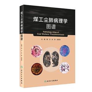
煤工尘肺病理学图谱(3) 版权信息
- ISBN:9787117310734
- 条形码:9787117310734 ; 978-7-117-31073-4
- 装帧:一般胶版纸
- 册数:暂无
- 重量:暂无
- 所属分类:>
煤工尘肺病理学图谱(3) 内容简介
全书精选了约260幅煤工尘肺大体与显微镜下典型的病理形态学病变图像,按照病变发生、发展演变的规律进行了编著。在图谱导读部分对煤工尘肺病变的特点与演变规律进行了详细的阐述,并在图谱部分对每幅图像的病变特点又辅以中英文对照的图注说明。同时结合特殊染色将一些病变特点凸显得更加明晰。本书描绘了煤工尘肺肺部病变,同时也是研究疾病发生、发展变化规律的重要参考
煤工尘肺病理学图谱(3) 目录
图 谱 导 读
**部分 肺脏的基本结构与形态学特点
1 肺脏的解剖学/ 2
2 肺脏的组织形态学 /3
2.1 肺实质:导气部和呼吸部 /3
2.2 肺间质:肺的血管、淋巴管和神经组织/ 6
第二部分 煤工尘肺形态学特点
1 煤工尘肺大体形态学特点 /8
1.1 煤斑灶:包括胸膜煤斑灶和肺内煤斑灶/ 8
1.2 结节病变/9
1.3 块状纤维化 9
1.4 弥漫性间质纤维化/ 9
1.5 肺气肿病变/ 9
1.6 淋巴结的病变(多见肺门淋巴结的累及)/ 10
1.7 胸膜病变 /10
1.8 煤工尘肺伴发肺结核病/ 10
1.9 煤工尘肺伴发肺癌 /10
1.10 煤工尘肺伴发肺源性心脏病/ 10
2 煤工尘肺组织形态学特点/10
2.1 煤矽肺 /10
2.2 矽煤肺(矽肺) /12
2.3 煤肺 /13
2.4 伴发性病变/ 13
2.5 其他病变 /14
参考文献 /16
Atlas Guide
Part I Anatomy and Histology of Lung
1 Anatomy of the lung/ 20
2 Histology of the lung/21
2.1 Parenchyma: It includes conduction portion and respiratory portion / 21
2.2 Mesenchyma: It contains blood vessels, lymphatic vessels and nerves / 25
Part II Pathology of Coal Workers' Pneumoconiosis
1 Gross appearance of coal workers' pneumoconiosis/ 27
1.1 Coal dust maculae/27
1.2 Coal dust nodules /28
1.3 Massive fi brosis /28
1.4 Diffuse interstitial fi brosis / 29
1.5 Emphysema /29
1.6 Lesions in the lymph nodes (usually in hilar lymph nodes)/ 29
1.7 Lesions in pleura /29
1.8 Coal works' pneumoconiosis with tuberculosis / 29
1.9 Coal works' pneumoconiosis with lung cancer /30
1.10 Coal works' pneumoconiosis with cor pulmonale /30
2 Histological changes of coal workers' pneumoconiosis/ 30
2.1 Anthracosilicosis/ 30
2.2 Silicosis/32
2.3 Anthracosis/33
2.4 Complicated lesions / 34
2.5 Others /35
References /37
图谱部分Atlas
**部分 煤工尘肺大体形态学特点Part I Gross Appearance of CoalWorkers' Pneumoconiosis
图1 胸膜煤斑灶/43
Fig.1 Pleural coal dust maculae / 43
图2 至图4 肺内煤斑灶/43
Fig.2 to Fig.4 Pulmonary coal dust maculae / 43
图5 肺内煤斑灶伴大泡型肺气肿形成/ 45
Fig.5 Pulmonary coal dust maculae accompanied by bullous emphysema /45
图6 肺内煤斑灶伴囊泡型肺气肿形成/ 45
Fig.6 Pulmonary coal dust maculae with cystic emphysema / 45
图7 和图8 肺内煤斑灶(全肺大切片标本) / 46
Fig.7 and Fig.8 Pulmonary coal dust maculae(the large slice) / 46
图9 和图10 肺内煤斑灶(肺叶大切片标本) / 47
Fig.9 and Fig.10 Pulmonary coal dust maculae(the large slice)/ 47
图11 和图12 肺内煤斑灶、煤尘纤维灶(煤结节)形成/ 48
Fig.11 and Fig.12 Pulmonary coal dust maculae and fi brotic lesions( coal dust nodule) / 48
图13 和图14 肺内煤斑灶、煤矽结节/ 49
Fig.13 and Fig.14 Pulmonary coal dust maculae and coal silicotic nodules /49
图15 和图16 肺内煤斑灶、煤矽结节/ 50
Fig.15 and Fig.16 Pulmonary coal dust maculae and coal silicotic nodules/ 50
图17 和图18 肺内煤斑灶51
Fig.17 and Fig.18 Pulmonary coal dust maculae 51
图19 和图20 进行性块状纤维化/ 52
Fig.19 and Fig.20 Progressive massive fi brosis/ 52
图21 和图22 进行性块状纤维化(肺大切片标本) / 53
Fig.21 and Fig.22 Progressive massive fi brosis(the large slice) / 53
图23 和图24 结节融合型块状纤维化/ 54
Fig.23 and Fig.24 Nodular confl uent massive fi brosis / 54
图25 和图26 结节融合型块状纤维化(同图23 肺大切片标本) / 55
Fig.25 and Fig.26 Nodular confl uent massive fi brosis( this large slice shares the samplewith Fig.23)/55
图27 和图28 混合型块状纤维化/ 56
Fig.27 and Fig.28 Mixed massive fi brosis / 56
图29 至图38 煤工尘肺伴结核病/ 57
Fig.29 to Fig.38 Coal works' pneumoconiosis with tuberculosis / 57
图39 煤工尘肺伴肺癌(周围型) / 62
Fig.39 Coal works' pneumoconiosis with lung cancer( Peripheral type) / 62
图40 煤工尘肺伴肺癌(弥漫型) / 62
Fig.40 Coal works' pneumoconiosis with lung cancer( Diffuse type)/ 62
图41 和图42 煤工尘肺伴胸膜粘连与增厚/ 63
Fig.41 and Fig.42 Coal works' pneumoconiosis with pleural adhesion and thickening/ 63
图43 和图44 煤工尘肺合并肺源性心脏病/ 64
Fig.43 and Fig.44 Coal works' pneumoconiosis with cor pulmonale / 64
第二部分 煤工尘肺组织形态学特点
Part II Histological Appearance of
Coal Workers' Pneumoconiosis
图45 至图48 胸膜煤斑/66
Fig.45 to Fig.48 Coal dust dots of pleura/ 66
图49 至图52 肺内煤斑/68
Fig.49 to Fig.52 Coal dust dots of lung /68
图53 和图54 煤尘细胞灶70
Fig.53 and Fig.54 Cellular coal dust foci / 70
图55 至图58 煤尘纤维灶/71
Fig.55 to Fig.58 Fibrous coal dust foci / 71
图59 至图74 大小不等、形状各异的煤尘灶/ 73
Fig.59 to Fig.74 Coal dust lesions varying in size and shape / 73
图75 至图78 巨噬细胞性肺泡炎/ 77
Fig.75 to Fig.78 Macrophage alveolitis / 77
图79 至图82 煤尘细胞性结节/ 79
Fig.79 to Fig.82 Cellular coal dust nodules / 79
图83 至图86 细胞纤维性结节/ 81
Fig.83 to Fig.86 Cellular fi brous nodules /81
图87 至图94 煤矽结节/83
Fig.87 to Fig.94 Coal silicotic nodules / 83
图95 不典型煤矽结节/88
Fig.95 Atypical coal silicotic nodule / 88
图96 至图103 融合型煤矽结节/ 89
Fig.96 to Fig.103 Confl uent coal silicotic nodules/ 89
图104 矽结节/93
Fig.104 Silicotic nodule /93
图105 融合型矽结节/93
Fig.105 Confl uent silicotic nodules / 93
图106 至图109 矽结节/94
Fig.106 to Fig.109 Silicotic nodule/ 94
图110 至图113 进行性块状纤维化/ 96
Fig.110 to Fig.113 Progressive massive fi brosis / 96
图114 和图115 融合型块状纤维化 / 98
Fig.114 and Fig.115 Confl uent massive fi brous lesion / 98
图116 和图117 弥漫性尘性间质纤维化 / 99
Fig.116 and Fig.117 Diffuse coal interstitial fi brosis/ 99
图118 和图119 间质纤维化伴平滑肌细胞局灶性增生 / 100
Fig.118 and Fig.119 Interstitial fi brosis with local proliferated smooth muscle cells / 100
图120 和图121 煤工尘肺伴结核病变/101
Fig.120 and Fig.121 Coal workers' pneumoconiosis with tuberculosis / 101
图122 肺内早期煤矽结核结节/ 102
Fig.122 Early stage of coal silicotic tuberculous nodule / 102
图123 肺内典型的煤矽结核结节/ 102
Fig.123 Classic coal silicotic tuberculous nodule / 102
图124 肺内煤矽结核结节/103
Fig.124 Coal silicotic tuberculous nodules/ 103
图125 至图127 肺内融合型煤矽结核结节/103
Fig.125 to Fig.127 Confl uent coal silicotic tuberculous nodules / 103
图128 和图129 煤工尘肺矽结核结节/105
Fig.128 and Fig.129 Silicotic tuberculous nodules of coal works' pneumoconiosis / 105
图130 结节融合型块状纤维化/ 106
Fig.130 Nodular confl uent massive fibrosis / 106
图131 至图134 煤工尘肺伴播散性粟粒性肺结核/ 106
Fig.131 to Fig.134 Disseminated miliary tuberculosis of coal workers' pneumoconiosis / 106
图135 尘性结核性结节/108
Fig.135 Coal dust tuberculous nodules / 108
图136 和图137 尘性结核性肉芽肿(结节)/ 109
Fig.136 and Fig.137 Coal dust tuberculous granuloma( nodule) / 109
图138 和图139 尘性结核性支气管炎/ 110
Fig.138 and Fig.139 Coal dust tuberculous bronchiolitis / 110
图140 煤工尘肺伴结核性胸膜炎/ 111
Fig.140 Tuberculous pleuritis of coal workers' pneumoconiosis / 111
图141 煤工尘肺结核性空洞/ 111
Fig. 141 Tuberculous cavity of coal workers' pneumoconiosis . 111
图142 和图143 煤工尘肺伴发肺腺癌/ 112
Fig.142 and Fig.143 Coal workers' pneumoconiosis with pulmonary adenocarcinoma / 112
图144 和图145 煤工尘肺伴淋巴结转移腺癌/ 113
Fig.144 and Fig.145 Coal workers' pneumoconiosis with lymph node adenocarcinoma metastasis / 113
图146 和图147 煤工尘肺伴发肺腺癌/ 114
Fig.146 and Fig.147 Coal workers' pneumoconiosis with pulmonary adenocarcinoma / 114
图148 至图151 煤工尘肺伴发腺鳞癌/ 115
Fig.148 to Fig151 Coal workers' pneumoconiosis with pulmonary adenosquamous carcinoma / 115
图152 和图153 煤工尘肺伴发恶性间皮瘤/117
Fig.152 and Fig.153 Coal workers' pneumoconiosis with malignant mesothelioma/ 117
图154 煤工尘肺伴发恶性间皮瘤淋巴结转移/ 118
Fig.154 Coal workers' pneumoconiosis with lymph node malignant mesothelioma
metastasis 118
图155 血管内瘤栓形成118
Fig.155 Carcinoma cells emboli in small blood vessels / 118
图156 至图159 淋巴结煤尘沉积. 119
Fig.156 to Fig.159 Lymph nodes with coal dust deposition . 119
图160 至图163 淋巴结内煤矽结节. 121
Fig.160 to Fig.163 Coal silicotic nodules in lymph nodes / 121
图164 淋巴结病变/123
Fig.164 Silicotic lesions in lymph nodes/ 123
图165 至图167 淋巴结内矽结节/ 123
Fig.165 to Fig.167 Silicotic nodules in lymph node / 123
图168 至图171 淋巴结内融合型矽结节/ 125
Fig.168 to Fig.171 Confl uent silicotic nodules in lymph node / 125
图172 和图173 淋巴结内煤矽结核结节/ 127
Fig.172 and Fig.173 Coal silicotic tuberculous nodules in lymph nodes / 127
图174 和图175 淋巴结内矽结核结节. 128
Fig.174 and Fig.175 Silicotic tuberculous nodules in lymph nodes / 128
图176 至图180 煤尘侵及支气管/129
Fig.176 to Fig.180 Coal dust deposition in bronchi /129
图181 至图183 尘性慢性细支气管炎/ 131
Fig.181 to Fig.183 Coal chronic bronchiolitis /131
图184 至图189 胸膜病变/133
Fig. 814 to Fig.189 Pleural lesions / 133
图190 煤尘沉积在肺内小支气管/ 136
Fig.190 Coal dust deposition in small bronchia / 136
图191 煤尘沉积在肺内细支气管及终末细支气管/ 136
Fig.191 Coal dust deposition in small bronchia and terminal bronchia / 136
图192 和图193 煤尘沉积在呼吸性细支气管及其分支/ 137
Fig.192 and Fig.193 Coal dust deposition in respiratory bronchia and its branaches /137
图194 尘性小叶中心型肺气肿/138
Fig.194 Coal dust-induced centriacinar emphysema /138
图195 全小叶破坏型肺气肿/ 138
Fig.195 Destructive panacinar emphysema / 138
图196 和图197 累及肺动脉/ 139
Fig.196 and Fig.197 Lesions in pulmonary artery / 139
图198 肺内小动脉病变/140
Fig.198 Lesions in pulmonary small arteries / 140
图199 肺内小血管病变/140
Fig.199 Lesions in pulmonary small vessels / 140
图200 和图201 小动脉病变/141
Fig.200 and Fig.201 Lesions in small arteries / 141
图202 煤尘累及淋巴结及其脂肪组织/ 142
Fig.202 Coal dust deposits involve lymph nodes and adipose tissue / 142
图203 煤尘累及支气管及其周围脂肪组织/ 142
Fig.203 Involvement of bronchi and adjacent fat tissue by coal dusts / 142
图204 和图205 淋巴管病变/ 143
Fig.204 and Fig.205 Lesions in the lymphatic vessels / 143
图206 和图207 胸膜下神经纤维受累/144
Fig.206 and Fig.207 Coal dusts involve subpleural nerve fi bers / 144
图208 煤工尘肺伴浆液出血性炎/ 145
Fig.208 Coal workers' pneumoconiosis with serous hemorrhagic infl ammation / 145
图209 煤工尘肺伴肺水肿/145
Fig.209 Coal workers' pneumoconiosis with pulmonary edema / 145
图210 和图211 煤工尘肺伴肺淤血、肺水肿 /146
Fig.210 and Fig.211 Coal workers' pneumoconiosis with pulmonary congestion and edema/ 146
图212 煤工尘肺伴化脓性炎/ 147
Fig.212 Coal workers pneumoconiosis with purulent infl ammation / 147
图213 煤工尘肺伴脓肿/147
Fig.213 Coal workers' pneumoconiosis with abscess / 147
图214 煤工尘肺伴浆液、纤维素性炎/ 148
Fig.214 Coal workers' pneumoconiosis with serous fi brinous infl ammation / 148
图215 煤工尘肺伴浆液、化脓性炎/ 148
Fig.215 Coal workers' pneumoconiosis with serous purulent infl ammation / 148
图216 和图217 煤工尘肺“棒状小体”形成 /149
Fig.216 and Fig.217 “Rod-shaped” bodies of coal workers' pneumoconiosis / 149
图218 至图223 肌成纤维细胞分化/ 150
Fig.218 to Fig.223 Myofi broblasts differentiation / 150
图224 正常心肌组织(右心室)/ 154
Fig.224 Normal heart tissue( from right ventricle)/ 154
图225 肺源性心脏病心肌组织(右心室)/ 154
Fig.225 Right ventricle with cor pulmonale / 154
第三部分 煤工尘肺案例
Part III Cases of Coal Workers' Pneumoconiosis
案例一 煤矽肺Ⅰ期(结节+ 尘斑- 气肿) /156
Case 1 Anthracosilicosis(stage Ⅰ,Nodules and maculae surrounded by emphysema) / 157
案例二 煤肺Ⅰ期(煤尘斑灶+ 细胞性结节) / 161
Case 2 Anthracosis(stage Ⅰ,Coal dust maculae and cellular nodules) /162
案例三 煤矽肺Ⅱ期(尘斑- 气肿型) 166
Case 3 Anthracosilicosis( stage Ⅱ,Coal dust maculae and emphysema) / 167
案例四 煤矽肺Ⅲ期(大块纤维化) / 170
Case 4 Anthracosilicosis(stage Ⅲ,Massive fi brosis) / 171
案例五 煤矽肺Ⅲ期合并肺结核病/ 176
Case 5 Anthracosilicosis( stage Ⅲ) with pulmonary tuberculosis /177
案例六 煤矽肺Ⅲ期合并肺癌 /182
Case 6 Anthracosilicosis( stage Ⅲ) with lung cancer / 183
案例七 煤肺Ⅰ期(煤斑- 气肿)合并肺源性心脏 / 189
Case 7 Anthracosis( stage Ⅰ,Coal dust maculae and emphysema) with cor pulmonale /191
后记 /198
Acknowledgement/ 199
展开全部
煤工尘肺病理学图谱(3) 作者简介
杨方,华北理工大学教授,主任医师,博士生导师。河北省第十一届政协常委,唐山市第十届、第十一届政协副主席,民进唐山市委第六届、第七届主任委员。全国很好教师,河北省省管很好专家,河北省有突出贡献中青年专家,中国侨界杰出人物提名奖和贡献奖。曾任河北省医学会病理学分会副主任委员,河北省生理科学会副理事长,唐山市医学会病理学分会主任委员。发表论文260余篇(SCI收录60余篇),主编著作2部,参编著作1部。主持与完成国家自然科学基金项目3项,国家973课题(协作单位负责人)1项。省(部)、市(厅)级科研项目10余项。获河北省科技进步二等奖2项、三等奖1项,能源部、原卫生部科技进步三等奖各1项,中国煤炭工业科学技术三等奖1项,河北省教学成果三等奖1项。


